How your brain works
The brain and nervous system
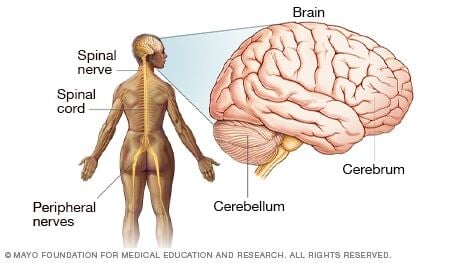
The brain contains billions of nerve cells arranged in patterns that coordinate thought, emotion, behavior, movement and sensation.
A complicated highway system of nerves connects the brain to the rest of your body, so communication can occur in seconds. Think about how fast you pull your hand back from a hot stove. While all the parts of the brain work together, each part is responsible for a specific function — controlling everything from your heart rate to your mood.
Cerebrum
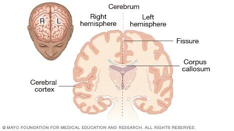
The cerebrum is the largest part of the brain. It's what you probably visualize when you think of brains in general. The outermost layer of the cerebrum is the cerebral cortex, also called the "gray matter" of the brain. Deep folds and wrinkles in the brain increase the surface area of the gray matter, so more information can be processed.
The cerebrum is divided by a deep groove, also known as a fissure. The groove divides the brain into two halves known as hemispheres. The hemispheres communicate with each other through a thick tract of nerves called the corpus callosum at the base of the groove. In fact, messages to and from one side of the body are usually handled by the opposite side of the brain.
Lobes of the brain
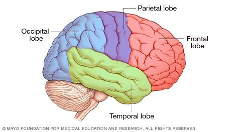
The brain's hemispheres have four lobes.
- The frontal lobes help control thinking, planning, organizing, problem-solving, short-term memory and movement.
- The parietal lobes help interpret feeling, known as sensory information. The lobes process taste, texture and temperature.
- The occipital lobes process images from your eyes and connect them to the images stored in your memory. This allows you to recognize images.
- The temporal lobes help process information from your senses of smell, taste and sound. They also play a role in memory storage.
Cerebellum and brainstem
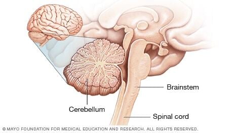
The cerebellum is a wrinkled ball of tissue below and behind the rest of the brain. It works to combine sensory information from the eyes, ears and muscles to help coordinate movement. The cerebellum activates when you learn to play the piano, for example.
The brainstem links the brain to the spinal cord. It controls functions vital to life, such as heart rate, blood pressure and breathing. The brainstem also is important for sleep.
The inner brain
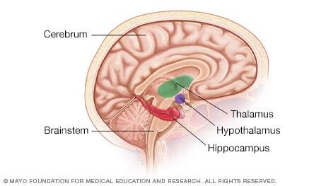
Structures deep within the brain control emotions and memories. Known as the limbic system, these structures come in pairs. Each part of this system is present in both halves of the brain.
- The thalamus acts as a gatekeeper for messages passed between the spinal cord and the cerebrum.
- The hypothalamus controls emotions. It also regulates your body's temperature and controls functions such as eating or sleeping.
- The hippocampus sends memories to be stored in areas of the cerebrum. It then recalls the memories later.
Peripheral nervous system
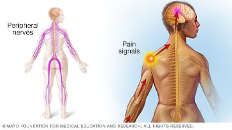
All of the nerves in your body that are outside of the brain and spinal cord make up the peripheral nervous system.
It relays information between your brain and your extremities, such as your arms, hands, legs or feet. For example, if you touch a hot stove, pain signals travel from your finger to your brain in a split second. Your brain tells the muscles in your arm and hand to quickly take your finger off the hot stove.
Nerve cells
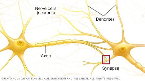
Nerve cells, known as neurons, send and receive nerve signals. They have two main types of branches coming off their cell bodies. Dendrites receive messages from other nerve cells. Axons carry outgoing messages from the cell body to other cells — such as a nearby neuron or muscle cell.
Interconnected with each other, neurons provide efficient, lightning-fast communication.
Neurotransmitters
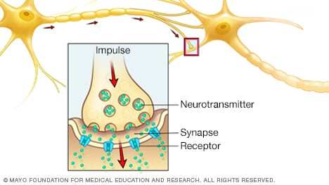
A nerve cell communicates with other cells through electrical impulses when the nerve cell is stimulated. Within a neuron, the impulse moves to the tip of an axon and causes the release of chemicals, called neurotransmitters, that act as messengers.
Neurotransmitters pass through the gap between two nerve cells, known as the synapse. They then attach to receptors on the receiving cell. This process repeats from neuron to neuron as the impulse travels to its destination. This web of communication that allows you to move, think, feel and communicate.


